When we consider the metabolic profile of healthy adult tissues, we find a highly standardized blueprint. Cells typically absorb glucose and, in the presence of oxygen, channel it through their mitochondria to maximize the production of adenosine triphosphate (ATP), the universal currency of cellular energy. This highly efficient process of oxidative phosphorylation is the default setting for almost all healthy organs.
However, a notable exception exists directly behind our pupils.
The Warburg Paradox in the Retina
One hundred years ago, the Nobel laureate Otto Warburg discovered a puzzling phenomenon. When analyzing the energy metabolism of the retina, he observed that even in the presence of abundant oxygen, this delicate neural tissue converts a vast majority of its glucose into lactate (lactic acid) rather than fully oxidizing it. At the time, this specific metabolic bypass—known as aerobic glycolysis, or the Warburg effect—had only been observed in one other type of tissue: rapidly multiplying, aggressive cancer cells.
To find this same metabolic signature in a healthy, highly developed sensory organ was a profound biological paradox. Today, modern biochemistry is revealing that the retina's cancer-like metabolism is not a flaw, but an example of cooperative biological engineering.
The Purpose Behind the Bypass
At first glance, opting for glycolysis over mitochondrial oxidation seems like an inefficient choice. Glycolysis yields only a fraction of the ATP that a mitochondrion can produce from a single molecule of glucose. Why would the retina, which is among the most metabolically active and energy-demanding tissues in the human body, rely on a less efficient pathway?
The answer lies in the intense physical demands of sight.
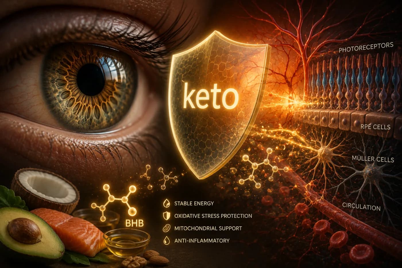
Photoreceptors—the specialized rods and cones that capture light—operate under continuous, high-stakes cellular stress. Exposed to intense light and high concentrations of oxygen, the delicate lipid membranes in their outer segments suffer rapid oxidative damage. To prevent permanent blindness, these outer segments must undergo constant, rapid renewal.
Every single day, the Retinal Pigment Epithelium (RPE), a supporting cell layer directly behind the photoreceptors, must digest and recycle approximately 10% of the photoreceptors' light-sensing outer segments. To keep pace with this rigorous destruction, the photoreceptors must continuously synthesize new proteins, lipids, and nucleic acids to rebuild themselves from scratch.
This is where the Warburg effect becomes essential. If the retina fully oxidized all of its glucose into carbon dioxide and water to produce maximum energy, it would leave the cells with plenty of ATP but no raw physical building blocks. By stopping the metabolic process early at the stage of glycolysis, the retina preserves the carbon skeletons of glucose. These carbon rings and chains are diverted into biosynthetic pathways, acting as the essential bricks and mortar needed to manufacture new cellular membranes.
In short, the retina performs aerobic glycolysis not because it lacks power, but because it is constantly rebuilding its physical structure.
A Cooperative Ecosystem of Energy
This metabolic choice requires a highly coordinated partnership between the different cell types of the eye. The retina does not work in isolation; it functions as a cooperative metabolic ecosystem where resources are carefully partitioned.
The photoreceptors and supporting glial cells, such as Müller glia, work in harmony to manage this delicate fuel pipeline. The Müller glia absorb glucose from the blood supply and perform glycolysis, producing lactate. This lactate is not treated as waste; instead, it is exported and absorbed by the photoreceptors, which can use it as a highly efficient fuel source.
Meanwhile, the retina still retains a high concentration of mitochondria. These cellular powerhouses are not scattered randomly; instead, they are tightly packed into a specialized region of the photoreceptor called the ellipsoid, situated immediately adjacent to the light-sensitive machinery. This strategic arrangement ensures that the high energy required to operate the ion pumps of the visual system is generated exactly where it is needed most, while the surrounding cytoplasm remains focused on biosynthetic renewal.
When the Balance Fails: Metabolic Disease and Sight
Because the retina's metabolism operates on such a tight and demanding wire, even minor disruptions to this delicate balance can have severe consequences for our vision.
The high-velocity blood supply of the choroid and the complex cellular maintenance pathways inside the eye must operate perfectly to keep this system stable. When mitochondria begin to decline due to aging or genetic factors, the retina's ability to balance energy production and structural repair is compromised.
Many major blinding diseases are now understood to have deep metabolic roots:
Age-Related Macular Degeneration (AMD). Impairments in mitochondrial function and a breakdown in the RPE's recycling process can lead to the accumulation of toxic cellular waste, slowly degrading central vision.
Diabetic Retinopathy. Fluctuations in systemic blood glucose disrupt the delicate glycolytic balance of the retina, damaging the microscopic capillaries that sustain this high-energy tissue.
By studying how healthy cells manage these massive metabolic demands, researchers are finding new ways to protect vision. Using advanced imaging technologies, such as two-photon microscopy, scientists can now monitor ATP and lactate levels within individual cells in real time, opening up new possibilities for detecting and treating metabolic eye diseases long before structural damage occurs.
Appreciating the Living Engineering
We often view our eyes as elegant optical instruments, comparing them to high-resolution cameras. But a camera is a static object made of glass and plastic.
Our visual system, by contrast, is a living, breathing example of high-energy engineering. To give us a seamless view of the world, our retinas continuously balance the destructive forces of light and oxygen with an equally powerful, cancer-like engine of biochemical reconstruction. The very metabolic process we associate with disease is, in the unique environment of the eye, the secret to preserving our window on the world.