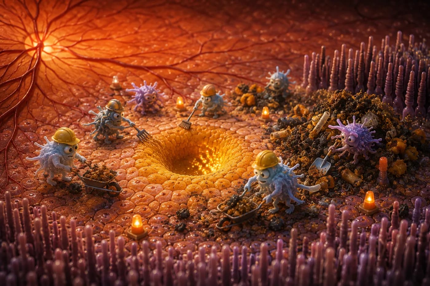
When we talk about vision loss and aging, we frequently encounter the term "Age-Related Macular Degeneration" (AMD). For most people, it sounds like an abstract, inevitable clinical diagnosis that comes with growing older. Yet, if we zoom into the retina under a microscope, we find a literal molecular battleground.
This is a dynamic ecosystem where specialized support cells and resident immune guards fight daily to keep our visual pathways clear of toxic waste. When this delicate balance fails, our visual architecture collapses.
Understanding how this biological waste disposal system operates—and what happens when it becomes chronically clogged—gives us the power to actively protect our sight. It is not an inevitable decline, but a process we can directly influence.
The Unsung Heroes: The Retinal Pigment Epithelium
At the core of vision are photoreceptors—the light-sensing rods and cones. They capture photons and transmit electrical signals to the visual cortex of the brain to assemble the images we see. These cells operate under extreme metabolic stress and cannot be replaced.
Because they are constantly bombarded by intense light and reactive oxygen, their sensitive outer segments become heavily damaged and oxidized daily. To survive, photoreceptors perform a brilliant biological trick: every morning, as the sun rises, they shed their worn-out, oxidized tips.
Directly beneath these photoreceptors lies a single layer of cells called the Retinal Pigment Epithelium (RPE). The RPE cells act as the eye's dedicated recycling stations. Their primary job is to engulf, digest, and recycle this massive daily pile of outer segment waste, returning critical nutrients back to the photoreceptors.
RPE cells are some of the most active and hardworking phagocytic cells in the entire human body. If they fail to clear the debris, our visual field quickly becomes clouded by metabolic waste.
When the Cleaners Tire: Microglia and the TREM2 Rescue Mission
As we age, our RPE cells naturally begin to slow down. Their structural framework stiffens, they decrease in number, and their lysosomal machinery struggles to process the daily waste load. When debris starts to accumulate, the body deploys an emergency backup crew: microglia, the specialized immune cells of the central nervous system.
In a healthy young eye, microglia reside strictly in the inner layers of the retina. However, during aging and RPE exhaustion, they translocate directly into the subretinal space—the tight junction between RPE cells and photoreceptors. This is a vital rescue mission.
The subretinal microglia step in to assist the overworked RPE, actively engulfing accumulated lipids and cellular debris to preserve visual acuity.
At the heart of this rescue mission is a molecular sensor on the microglial surface known as the TREM2 receptor. TREM2. Acts as a lipid scanner. It detects accumulated toxic fats and cellular waste, triggering the microglia to engulf and degrade them. When TREM2 signaling is healthy, the retina resists age-related decay and maintains sharp vision.
A Double-Edged Sword: When Protectors Turn Destructive
However, under conditions of chronic systemic inflammation, poor diet, and sedentary lifestyles, this protective immune response undergoes a pathological shift. The microglia that rushed to save the retina become hyperactive and destructive, leading directly to the two main forms of AMD.
Dry AMD and the Demolition of the Barrier (SPP1+ Microglia)
Under prolonged oxidative stress, a specific subgroup of disease-associated microglia (DAM) emerges in the subretinal space, releasing a protein called SPP1. This protein forces RPE cells to undergo Epithelial-Mesenchymal Transition (EMT).
As a result, the vital RPE cells lose their hexagonal shape, and their crucial tight junctions (proteins like ZO-1 and claudin-1)—which form the blood-retinal barrier—disintegrate. The neat, protective RPE monolayer collapses into disorganized cell clumps.
Without this essential barrier, photoreceptors are left unprotected and quickly die, leading to Dry AMD and Geographic Atrophy.
Wet AMD and Pathological Neovascularization
When activated microglia remain trapped in the subretinal space long-term, they stimulate RPE cells to secrete high levels of pro-inflammatory cytokines and vascular endothelial growth factor (VEGF), alongside tissue-destroying enzymes known as matrix metalloproteinases (MMP1, MMP2, MMP9).
These enzymes degrade Bruch's membrane, the thin barrier separating the retina from the blood vessels beneath it. Fragile, abnormal blood vessels begin to sprout through these micro-tears, leaking blood and fluid directly under the retina.
This drives Wet AMD, which can destroy central, high-resolution vision in a matter of weeks.
How to Support Your Eye's Cleaning Crew
The molecular battle in your retina is not a pre-determined outcome. Science demonstrates that we can directly support our RPE and microglial cells through two highly effective lifestyle habits:
Voluntary Aerobic Exercise. Rigorous studies show that regular, voluntary physical activity (such as self-paced running or brisk walking) rejuvenates the retinal transcriptome. It downregulates pro-inflammatory microglial genes and suppresses the complement cascade that degrades synapses. Crucially, the exercise must be voluntary; forced, stressful regimens do not produce the same genomic benefits.
Intermittent Fasting. Because visual cells run on an extremely high metabolic budget, constant digestion leaves their recycling engines overworked. Intermittent fasting stimulates autophagy—the cell's natural self-cleaning process. This gives RPE lysosomes the window they need to clear out lipid droplets and misfolded proteins before they aggregate into toxic, vision-blocking drusen.
At KSA Vision Clinic, we view vision as a living biological ecosystem. By choosing to prioritize deep sleep, voluntary exercise, and intentional fasting, you are directly signaling your genome to preserve your visual pathways, ensuring brilliant, crystal-clear vision for a lifetime.