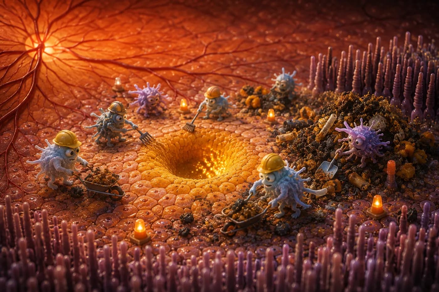
When we sit in a dark room with our eyes closed, our conscious mind tends to perceive this as a period of absolute rest. It is natural to assume that when there is no light to capture, the sensory pathways of the eye are simply paused, waiting for the morning.
However, cellular biology reveals a different and highly cooperative process.
The Active Visual System in Darkness
Beneath the surface, the visual system is active and highly coordinated. The retina, which is a developmental extension of the central nervous system, contains more than 100 million photoreceptor cells that are working continuously. Rather than resting in the absence of light, these cells consume oxygen and nutrients at an exceptionally high rate.
In terms of biological energetics, maintaining darkness is actually one of the most demanding tasks the visual system performs. This continuous activity is often described by researchers as the "dark current"—an elegant mechanism designed to keep our eyes in a state of constant, immediate readiness to perceive our surroundings.
Understanding the Dark Current
To understand why darkness requires so much energy, it is helpful to look at how rod and cone photoreceptors operate at a cellular level.
In many sensory systems, a receptor cell remains quiet until it is stimulated. For example, a touch receptor in the skin waits for physical contact, and a hair cell in the inner ear waits for sound waves. When stimulated, these cells depolarize, creating an electrical signal that travels to the brain.
Photoreceptors, however, function in the opposite manner. In absolute darkness, rod and cone cells are in a relatively depolarized state, maintaining a steady membrane potential of approximately -40 millivolts. To maintain this potential, the cell keeps cyclic guanosine monophosphate (cGMP) levels high, which holds specialized sodium and calcium channels open. This continuous influx of positive ions is the "dark current".
Because these channels are open in the dark, the cell is continuously active, releasing a steady stream of the neurotransmitter glutamate onto neighboring retinal neurons.
To keep this current flowing without disrupting the delicate balance of ions within the cell, the photoreceptor relies on a high density of sodium-potassium adenosine triphosphatase (Na+-K+ ATPase) pumps. These pumps require a continuous supply of adenosine triphosphate (ATP), the primary energy currency of the cell, to move ions back across the membrane.
When a photon of light is absorbed by the visual pigment, it triggers a cascade of biochemical events. This cascade activates phosphodiesterase, which breaks down cGMP, causing the ion channels to close. As the influx of sodium and calcium stops, the membrane potential becomes more negative—a process called hyperpolarization. This hyperpolarization reduces the release of glutamate.
In this sense, light serves as an inhibitory signal that reduces the cell's neurotransmitter release. It is darkness that represents the active, energy-demanding state for these primary visual detectors.
The Supporting Vascular Infrastructure
Fueling this continuous metabolic demand requires an exceptionally rich blood supply. The photoreceptors are located in the outer layer of the retina, positioned directly adjacent to a dense network of blood vessels called the choroid.
The choroid has one of the highest blood-flow rates per gram of tissue weight in the human body. This vascular system is designed to provide a steady, generous supply of oxygen and glucose to the outer retina without interfering with our vision.
Because light must pass through the front of the eye and the inner layers of the retina to reach the photoreceptors, the central visual path must remain transparent and free of large, obscuring vessels. Positioning the highly vascular choroid behind the photoreceptor layer resolves this challenge, allowing light to arrive unobstructed while ensuring the cells have the metabolic support they require.
Processing Power in a Compact Space
The scale of this biological system is remarkable. Each human retina contains roughly 100 to 120 million rod cells, which are highly sensitive to low light, and approximately 6 million cone cells, which manage color and high-acuity daytime vision.
While it is tempting to compare these light detectors to the pixels of a digital camera, biological vision is far more sophisticated than a simple collection of passive sensors. The retina is complex neural tissue. Before any signal enters the optic nerve, a network of specialized cells—including bipolar, horizontal, and amacrine cells—processes and compares the electrical potentials across the retina.
These local computations allow the retina to identify edges, detect motion, and adapt to changes in contrast and illumination before the information leaves the eye. Ultimately, the signals from over 100 million photoreceptors are compressed and funneled into approximately 1 million axons that make up the optic nerve. The eye does not simply capture a static image; it processes, refines, and packages visual data in real time.
Vision as an Indicator of Systemic Health
Because the visual system depends on such a high level of metabolic support and vascular coordination, eye health is deeply integrated with the health of the entire body.
Several physiological systems work in harmony to support our visual pathways:
Vascular Health. Because of the high blood flow within the choroid, the microvasculature of the eye is sensitive to systemic circulation issues. Consistent, healthy blood pressure supports the gentle delivery of nutrients.
Metabolic Stability. Stable glucose metabolism is essential for the continuous energy requirements of the retina, as metabolic disruptions can impact the retinal blood vessels over time.
Rest and Recovery. The visual pathway extends deep into the brain, where large neural networks interpret visual signals. The brain relies on regular sleep and recovery to maintain its cognitive and metabolic health.
Surface Hydration. Even the simple act of blinking plays a significant role. Under normal conditions, humans blink approximately 15 to 20 times per minute, which stabilizes the tear film and maintains a clear optical surface. During focused screen use, our blink rate often decreases, which can lead to dry eyes and minor visual distortion.
These connections illustrate that maintaining clear vision is a cooperative effort involving multiple systems throughout the body.
A Direct Window into the Body
The transparent design of the eye provides a unique opportunity for preventive medicine. Because the cornea and lens are clear, the retina is one of the only areas of the human body where medical professionals can directly observe living blood vessels and neural tissue without surgery.
During a comprehensive eye examination, an optometrist or ophthalmologist can inspect the microvasculature of the retina and the health of the optic nerve. Because systemic conditions such as diabetes or hypertension often affect small blood vessels first, these exams can sometimes reveal early indicators of broader health changes before symptoms appear elsewhere in the body.
A Appreciative Perspective on Sight
We often think of sight as a passive experience, assuming the world simply appears before us when we open our eyes.
In reality, visual perception is an active, constructive process. The brain receives a highly compressed and processed stream of neural signals and combines them with previous experiences, eye movements, and balance information to create a stable, continuous view of our surroundings.
The next time you rest in a dark room, it is worth remembering the gentle, quiet work taking place at the back of your eyes. While you sleep, the photoreceptors are maintained by their steady dark current, keeping the visual system prepared and waiting to welcome the very next photon of light.
Sources: National Eye Institute (NEI) — How the Eyes Work & About the Eye. NCBI Bookshelf — "Facts and Figures Concerning the Human Retina" & "Phototransduction". Physiological Reviews — "Retinal physiology and circulation" and "The Multifunctional Choroid". University of Zurich — Lab for Retinal Cell Biology.