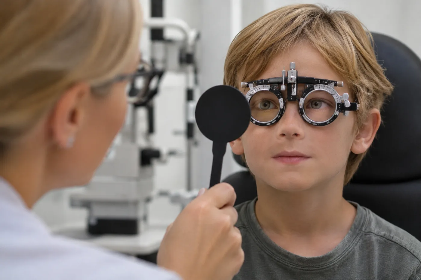
Did you know that the human body features a single, natural window through which doctors can directly observe living neural tissue and microvessels without making a single incision or injection? This window is not inside your skull—it is right inside your eyes.
When thinking about an eye clinic appointment, most people envision reading letters on a chart, getting fitted for contact lenses, or considering laser vision correction. We visit to check our visual sharpness. Yet groundbreaking discoveries in medical science reveal something far more profound: the retina at the back of your eye is not just a biological camera lens—it is an anatomical and functional extension of the central nervous system.
Today, neuroscientists and ophthalmologists worldwide are pursuing an inspiring question: could a routine eye scan help identify Alzheimer's disease years before the very first memory lapse appears?
The Brain's Outpost in the Eye
During embryonic development, the growing brain pushes out two small neuroectodermal buds that develop into our eyes. Consequently, the retina shares identical cell types, nerve fibers, microvascular networks, and immune mechanisms with cerebral brain tissue.
The greatest hurdle in combating Alzheimer's disease has always been lost time. The condition does not begin when someone misplaces their keys or forgets a familiar name. Scientists have proven that microscopic damage begins in the brain more than 15 years before clinical symptoms manifest. Today, diagnoses are often made only after substantial neural networks have been irreversibly lost.
However, if the retina is the brain's developmental "twin," do those same microscopic footprints appear in the eye? The scientific answer is yes.
What Postmortem Eye Studies Reveal Under the Microscope
Landmark histological and proteomic studies examining donor tissues from individuals with normal cognition, mild cognitive impairment (MCI), and Alzheimer's disease revealed striking evidence:
Protein Accumulation in the Retina. The primary drivers of Alzheimer's pathology are amyloid-beta plaques and abnormal tau proteins. Scientists discovered that amyloid deposits accumulate in the inner layers and margins of the retina as early as the mild cognitive impairment stage.
Ganglion Cell Loss. Alongside protein deposits, retinal ganglion cells and axonal fiber layers undergo progressive thinning and loss.
Inflammatory Activation. Retinal microglial cells activate in a pattern that directly mirrors the neuroinflammatory cascade occurring within the cerebral cortex.
Because the retina reflects cerebral pathology so accurately, researchers asked: can we detect these micro-changes in living patients safely, non-invasively, and affordably?
Measuring Cells with Light: What Is OCT?
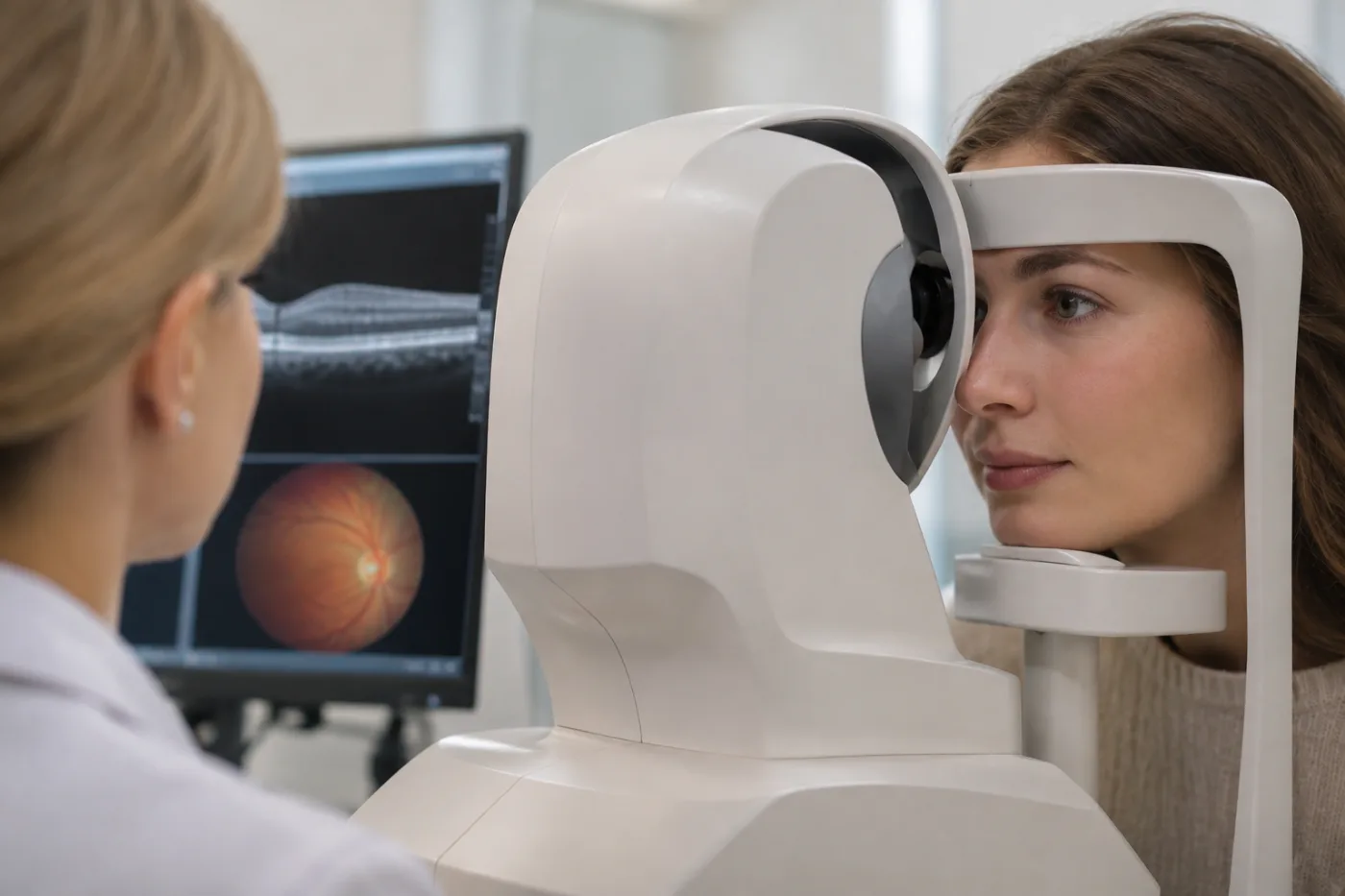
Many patients visiting modern eye centers are familiar with Optical Coherence Tomography (OCT). The exam is simple and comfortable: you sit in a chair, rest your chin on a support, look at a small light target, and within seconds the scanner captures a high-resolution, three-dimensional cross-section of your retina.
OCT uses completely safe light waves rather than ionizing radiation, requiring no needles and no physical contact with the eye. It measures retinal layers with micron-level precision (a human hair is roughly 70 microns thick; OCT measures down to individual microns!).
Advanced diagnostic algorithms analyze key inner retinal layers:
- RNFL (Retinal Nerve Fiber Layer)
- GCL (Ganglion Cell Layer)
- IPL (Inner Plexiform Layer)
In a landmark collaborative study analyzing health data from nearly 70,000 individuals across four cohorts (including the UK Biobank), researchers confirmed that individuals with thinner macular nerve fiber and ganglion cell layers exhibited a significantly higher risk of developing incident dementia over long-term follow-up. Retinal thinning served as a measurable signal of declining neural density.
The Synergy Between Eye Scans and Blood Tests
Historically, evaluating Alzheimer's risk required expensive PET imaging (costing thousands of euros with radioactive tracers) or invasive spinal taps to collect cerebrospinal fluid.
Today, medicine has made a breakthrough with high-sensitivity blood biomarkers, including p-tau217 and the neuroinflammation marker GFAP.
Recent clinical studies demonstrate that structural thinning of inner retinal layers on OCT directly mirrors rising levels of these plasma biomarkers in preclinical individuals. As inflammatory GFAP or pathological p-tau217 begins to increase in the bloodstream, the inner retinal layers simultaneously begin to thin before cognitive decline occurs. Combining non-invasive retinal imaging with accessible blood assays could soon provide a scalable, early-detection screening model.
Can an Eye Specialist Diagnose Alzheimer's Today?
To be transparent and scientifically responsible: not quite yet.
Retinal thinning can also stem from common eye conditions such as glaucoma or high myopia. Therefore, an isolated thin layer on an OCT scan is not a standalone diagnosis of dementia. Global research teams are currently training specialized artificial intelligence models to reliably distinguish between ocular and neurological patterns.
Nevertheless, the trajectory is clear: eye centers are emerging as vital frontlines for preventative health and longevity.
How You Can Check Your Retinal Health Today
While global researchers finalize clinical screening protocols for neurodegeneration, state-of-the-art retinal scanning technology is already an essential part of modern preventative eye care.
At KSA Eye Center (KSA Silmakeskus), high-precision OCT scans are standardly included within our comprehensive Silmade Audit (Eye Audit). The exam takes only minutes, is completely contactless and painless, and provides our optometrists and eye doctors with an in-depth, micron-level assessment of your retina, optic nerve, and corneal structure.
Undergoing a comprehensive Eye Audit not only confirms whether you have sharp vision or are a great candidate for laser vision correction—it also provides peace of mind by keeping a close watch on the underlying health of your visual and neural structures.
Take care of your vision—because caring for your eyes is caring for your mind!