Imagine looking through a pair of binoculars where the left lens is crystal clear, but the right lens is coated in a thick, stubborn fog. This is not science fiction—it is the daily visual reality for millions of people living with unequal optical power in their eyes. In the medical world, this condition is called anisometropia. Remarkably, when a child develops it, they almost never complain or mention that anything is wrong.
Parents are accustomed to hearing their children speak up about everyday discomforts: tight shoes, an itchy sweater, or a scraped knee. Yet rarely does a parent hear: "Mom, Dad, my left eye sees the world much blurrier than my right."
Why does this happen? What takes place within the brain when our eyes project two mismatched images, and why can conventional eyeglasses sometimes make the problem worse? Let's explore this fascinating intersection of optics, ophthalmology, and neuroscience in plain language.
What Is Anisometropia?
Under normal conditions, our eyes develop as a balanced pair: if a person is nearsighted or farsighted, the refractive error in both eyes is typically very similar.
Anisometropia (derived from the Greek an-iso-metron, meaning "unequal measurements") describes a condition where the refractive power of the two eyes differs significantly. Optometrists and ophthalmologists clinically diagnose anisometropia when the difference between the eyes is 1.0 diopter or greater. In clinical practice, however, differences of 3, 5, or even 8 diopters are not uncommon.
It can manifest in several distinct patterns:
- Simple Anisometropia: One eye has perfect, 20/20 vision, while the other eye has significant nearsightedness (myopia) or farsightedness (hyperopia).
- Antimetropia: A unique scenario where one eye is nearsighted (minus power) while the fellow eye is farsighted (plus power).
- Compound Anisometropia: Both eyes share the same type of refractive error, but one is significantly more pronounced than the other (e.g., -1.50 D in the right eye versus -6.00 D in the left eye).
The Silent Condition: Why Children Never Complain
One of the most perplexing moments for parents occurs during a preschool or school vision screening: a child who performs well academically, catches balls easily, and draws detailed pictures is suddenly found to have virtually no functional vision in one eye.
The reason lies in the brain's astonishing ability to compensate. Human beings navigate the world with binocular vision—both eyes open simultaneously. When a young child's visual cortex receives one crisp, sharp image from the stronger eye and one blurry, unfocused image from the weaker eye, the brain performs an automatic, brilliant shortcut: it simply processes the clear image and disregards the blur.
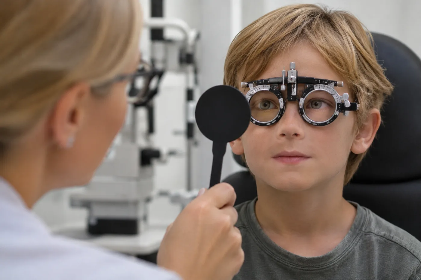
Because the child has never known any other way of seeing, their world appears completely normal! Only when a doctor or teacher covers the "good" eye does the child suddenly realize they cannot clearly distinguish objects with their fellow eye.
The Amblyopia Trap: Understanding the "Lazy Eye"
If childhood anisometropia remains undetected and uncorrected during the early formative years, it often leads to amblyopia, commonly known as "lazy eye."
To grasp why this happens, we must remember a fundamental truth: we see with our brains, not just our eyes. The eyes serve as optical lenses capturing light rays, but it is the visual cortex at the back of the brain that decodes these impulses into three-dimensional perception.
At birth, the neural wiring of the visual cortex is immature. It develops and refines its complex neural connections throughout the first 7 to 9 years of life. For this neural development to occur properly, the brain requires equal, high-definition visual signals from both eyes.
When one eye persistently delivers a defocused image, the visual cortex actively suppresses it to prevent double vision and visual confusion—a phenomenon known as cortical suppression. Deprived of stimulation, the neural pathways corresponding to the weaker eye stop developing.
If left untreated past the critical developmental window, the condition becomes permanent: even with corrective lenses or surgical procedures in adulthood, the eye cannot achieve 20/20 clarity because the brain's processing circuitry was never fully wired.
An Optical Puzzle: What Is Aniseikonia?
Once anisometropia is diagnosed, the intuitive solution seems straightforward: prescribe eyeglasses with different lens powers. However, this is where optics and physics introduce a secondary complication known as aniseikonia.
Aniseikonia occurs when the perceived image sizes on the two retinas are significantly different:
- Plus lenses (for farsightedness) magnify the retinal image like a magnifying glass.
- Minus lenses (for nearsightedness) minify the retinal image, making objects appear smaller.
As a general clinical rule, every diopter of power difference between spectacle lenses creates roughly a 1% to 2% difference in image size. With a 4-diopter discrepancy, one eye perceives an object 5% to 8% larger than the other.
When the brain attempts to fuse two identical objects of noticeably different scale into a single image, it experiences intense strain. Patients often experience:
- Frequent headaches and ocular fatigue (asthenopia);
- Disrupted stereoscopic depth perception;
- Spatial distortion, dizziness, or mild double vision.
Why Contact Lenses Often Provide a Superior Optical Solution
Eyeglass lenses sit approximately 12 to 14 millimeters in front of the cornea, and this vertex distance dramatically amplifies image magnification differences.
Contact lenses, by contrast, rest directly on the surface of the cornea—at the optical entrance of the eye. By eliminating this vertex distance, contact lenses reduce retinal image size disparities to minimal, comfortable levels (typically under 1%). The brain can comfortably fuse the two images into a cohesive, stereoscopic picture. For teens and adults with significant refractive differences, contact lenses often provide a far more comfortable, distortion-free visual experience than glasses.
Reassurance and Perspective: Thriving in an Active World
If you or an adult family member has anisometropia or longstanding amblyopia, there is every reason for optimism.
The human brain adapts remarkably well. Individuals with one dominant eye lead vibrant, active, and fully capable lives. They drive cars, play sports, excel in demanding professions, and navigate the physical world with ease. The brain readily utilizes monocular depth cues—such as motion parallax, shadows, perspective, and relative sizing—to judge distance accurately.
For young children, however, the call to action remains clear: during the critical neuroplastic window (up to age 8 or 9), early intervention can work wonders. When unequal refractive error is identified early and treated with proper optical correction and patching therapy, we can stimulate the weaker eye, restore neural connectivity, and give the child the lifelong gift of balanced binocular vision.
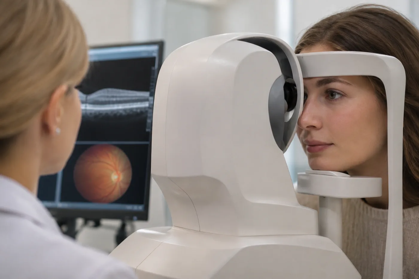
Protecting Your Vision Through Advanced Diagnostic Care
Because anisometropia is completely invisible to the naked eye, relying on outward appearances or waiting for a child to complain is never enough.
Prioritizing your visual health today ensures comfort, clarity, and peace of mind for a lifetime of vibrant sight!